
Epistemis

FISH · Cardiovascular · open case 08

Histology · Cardiovascular
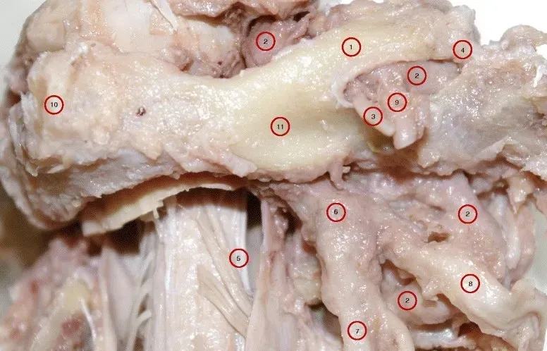
A higher magnification of specimen A of the right posterolateral aspect of sub-occipital region showing the atlas with complete dorsal and lateral bridges; and the oval opening between the two bridges. Also, the passage of vertebral artery through the tunnel formed by the posterior and lateral bridges. The sub-occipital nerve (C1 dorsal ramus) along with branches of VA emerging from the oval opening. 1. Dorsal bridge; 2. Vertebral artery; 3. C1 dorsal ramus; 4. Lateral bridge; 5. Denticulate ligament; 6. C2 dorsal root ganglion; 7. C2 dorsal ramus; 8. C2 ventral ramus; 9. Branches of VA; 10. Dorsal tubercle of atlas; 11. Dorsal arch of atlas.
A higher magnification of specimen A of the right posterolateral aspect of sub-occipital region showing the atlas with complete dorsal and lateral bridges; and the oval opening…

The guided interpretation is available in the app

Epistemis explains what is seen in this image, why, and what it should not be confused with: guided findings, reasoned interpretation, and the clinical pearls of the case. Free; The image becomes available offline after downloading.

Download Epistemis

Epistemis is educational review material. It is not a medical device, does not diagnose or prescribe treatment, and does not replace formal medical training, current clinical guidelines, or professional clinical judgment.

Category
Cardiovascular
Difficulty
advanced
Type
Applied histology
Mode
FISH
Stain
FISH

Origin: MultiCaRe · figure from open-access article PMC4724954 · License CC BY

Download Epistemis