
Epistemis

H&E · Skin · open case 02

Pathological anatomy · Skin
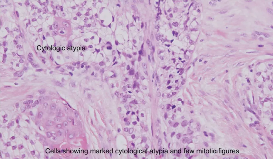
(A, B). High power The biopsy specimen showed marked proliferation of irregular sebaceous lobules of different sizes and shapes in the dermis. The cells of the lobules are mostly undifferentiated and demonstrated marked cylologic atypia with considerable nuclear pleomorphism and few mitotic activities. Some of the sebaceous lobules showed areas composed of atypical keratinizing cells. The overlying epidermis is normal with marked dermal fibrosis. Immunoperoxidase staining showed positive reaction for cytokeratin (CK), focal positive for epithelial membrane antigen (EMA) and negative reaction for carcino-embryonic antigen (CEA).
(A, B). High power The biopsy specimen showed marked proliferation of irregular sebaceous lobules of different sizes and shapes in the dermis. The cells of the lobules are mostly…

The guided interpretation is available in the app

Epistemis explains what is seen in this image, why, and what it should not be confused with: guided findings, reasoned interpretation, and the clinical pearls of the case. Free; The image becomes available offline after downloading.

Download Epistemis

Epistemis is educational review material. It is not a medical device, does not diagnose or prescribe treatment, and does not replace formal medical training, current clinical guidelines, or professional clinical judgment.

Category
Skin
Difficulty
advanced
Type
Clinical microscopy
Mode
H&E
Stain
H&E

Origin: MultiCaRe · figure from open-access article PMC3161692 · License CC BY

Download Epistemis