
Epistemis

H&E · Nervous system · open case 07

Pathological anatomy · Nervous system
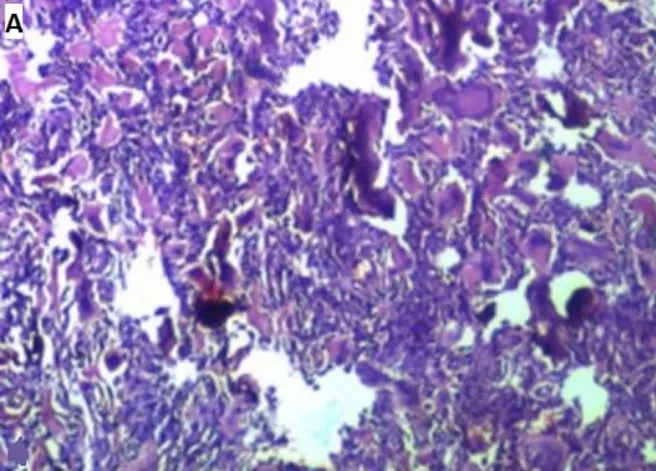
Microscopic histopathological images showing a focal necrosis tumor proliferation, vaguely lobulated and crossed by fibrous septa (H & E 400x); this proliferation is of moderate cellularity made up essentially of small round monomorphic basophilic cells with a rounded or oval nucleus with a high nuclear/cytoplasmic ratio; focally, there was a transition between these undifferentiated embryonic cells with other neurocytically differentiated cells; the background of the proliferation is clearly fibrillary with presence of numerous Homer-Wright rosette images and pseudo-rosettes (H & E 400x); it is associated with micro calcifications and rare spumous histiocytes.
Microscopic histopathological images showing a focal necrosis tumor proliferation, vaguely lobulated and crossed by fibrous septa (H & E 400x); this proliferation is of moderate…

The guided interpretation is available in the app

Epistemis explains what is seen in this image, why, and what it should not be confused with: guided findings, reasoned interpretation, and the clinical pearls of the case. Free; The image becomes available offline after downloading.

Download Epistemis

Epistemis is educational review material. It is not a medical device, does not diagnose or prescribe treatment, and does not replace formal medical training, current clinical guidelines, or professional clinical judgment.

Category
Nervous system
Difficulty
advanced
Type
Clinical microscopy
Mode
H&E
Stain
H&E

Origin: MultiCaRe · figure from open-access article PMC8571934 · License CC BY

Download Epistemis