
Epistemis

Immunostaining · Hematolymphoid · open case 16

Pathological anatomy · Hematolymphoid
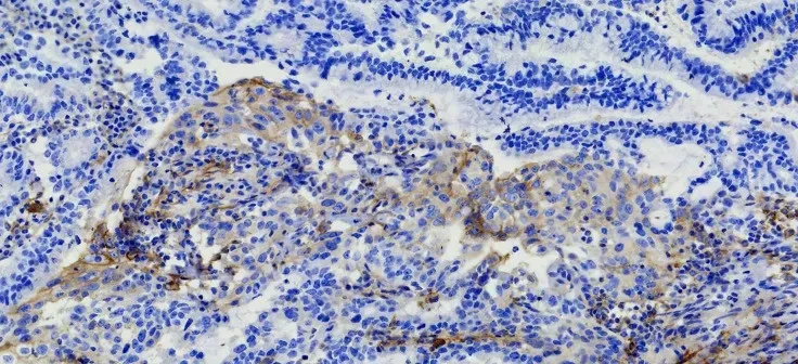
Immunohistochemical PD-L1 staining (Magnification: 10 x 40). The patient's expression level of PD-LI was detected using the VENTANA PD-L1 (SP263) Assay. The brown cells in the immunohistochemical staining for PD-L1 are positive cells, including tumor cells (TCs) with membrane staining at any intensity and immune cells (IC) in the tumor area, such as lymphocytes and macrophages. After counting the positive cells, the corresponding proportion was calculated according to the formula. Tumor Proportion Score (TPS), which is the percentage of viable tumor cells showing partial or complete membrane staining at any intensity. Combined positive score (CPS) = (positive TC + positive IC)/total tumor cells x 100.
Immunohistochemical PD-L1 staining (Magnification: 10 x 40). The patient's expression level of PD-LI was detected using the VENTANA PD-L1 (SP263) Assay. The brown cells in the…

The guided interpretation is available in the app

Epistemis explains what is seen in this image, why, and what it should not be confused with: guided findings, reasoned interpretation, and the clinical pearls of the case. Free; The image becomes available offline after downloading.

Download Epistemis

Epistemis is educational review material. It is not a medical device, does not diagnose or prescribe treatment, and does not replace formal medical training, current clinical guidelines, or professional clinical judgment.

Category
Hematolymphoid
Difficulty
advanced
Type
Clinical microscopy
Mode
Immunostaining
Stain
Immunostaining

Origin: MultiCaRe · figure from open-access article PMC7604326 · License CC BY

Download Epistemis