
Epistemis

Doppler · Neuro / Head · open case 006

Radiology · Neuro / Head
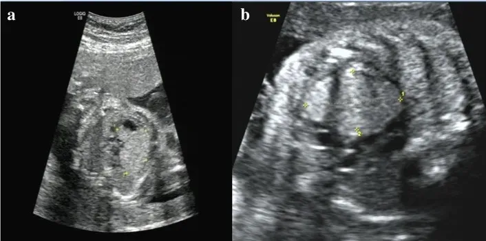
Color Doppler ultrasound imaging. a on November 3, 2017 (23 + 4 weeks of gestation), color Doppler ultrasound imaging showed a hyperechogenic mass in the fetal left ventricle, measuring 1.8 cm x 1.57 cm, broadening of the left lateral ventricle (1.11 cm) and a strong dot-like echo in the left ventricle; b on December 7, 2017, color Doppler ultrasound reexaminations displayed multiple strong echoes in the fetal left ventricle (measuring 3.3 cm x 2.03 cm), compression of the left ventricular outflow tract, obvious enlargement of the tumor, and a 0.92 cm internal diameter of posterior horn of the left lateral ventricle.
Color Doppler ultrasound imaging. a on November 3, 2017 (23 + 4 weeks of gestation), color Doppler ultrasound imaging showed a hyperechogenic mass in the fetal left ventricle,…

The guided interpretation is available in the app

Epistemis explains what is seen in this image, why, and what it should not be confused with: guided findings, reasoned interpretation, and the clinical pearls of the case. Free; The image becomes available offline after downloading.

Download Epistemis

Epistemis is educational review material. It is not a medical device, does not diagnose or prescribe treatment, and does not replace formal medical training, current clinical guidelines, or professional clinical judgment.

Category
Neuro / Head
Difficulty
mean
Type
Radiological image
Mode
Doppler

Origin: MultiCaRe · figure from open-access article PMC6537215 · License CC BY

Download Epistemis