
Epistemis

PET · Whole body · open case 021

Radiology · Whole body
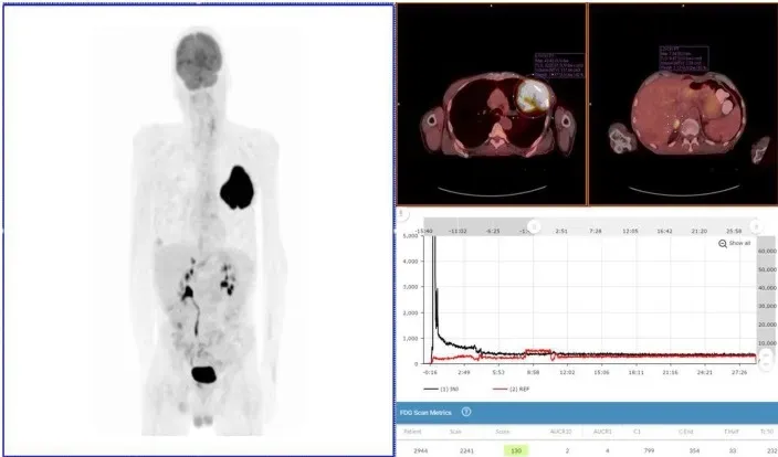
Exam 2:maximum intensity projection image, fused axial images, and time-activity curves of repeat PET/CT Scan. Repeat PET/CT MIP image (above left image), taken 3 days after first PET/CT and 65 min post-injection, reveals lung mass, right adrenal lesion, prostate lesion, and some minimal abnormal uptake in left forearm (radiotracer injection site was left hand). Black time-activity curve (lower right image) from injection arm sensor reveals a nearly ideal injection. Red time-activity curve (also lower right image) from contralateral arm sensor reveals expected uptake. Fused PET/CT images (upper right images) reveal left lobe mass (SUVmax 43) and adrenal lesion.
Exam 2:maximum intensity projection image, fused axial images, and time-activity curves of repeat PET/CT Scan. Repeat PET/CT MIP image (above left image), taken 3 days after first…

The guided interpretation is available in the app

Epistemis explains what is seen in this image, why, and what it should not be confused with: guided findings, reasoned interpretation, and the clinical pearls of the case. Free; The image becomes available offline after downloading.

Download Epistemis

Epistemis is educational review material. It is not a medical device, does not diagnose or prescribe treatment, and does not replace formal medical training, current clinical guidelines, or professional clinical judgment.

Category
Whole body
Difficulty
advanced
Type
Radiological image
Mode
PET

Origin: MultiCaRe · figure from open-access article PMC5962786 · License CC BY

Download Epistemis