
Epistemis

PET · Pelvis · open case 030

Radiology · Pelvis
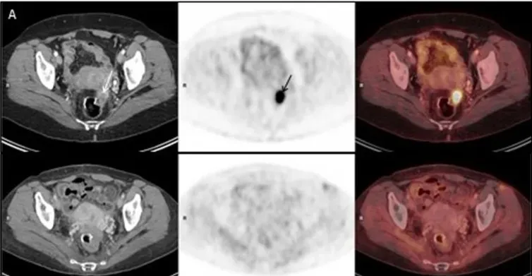
(A,B) represent PET-CT images of two different pelvic metastases in the radiation field. Upper panel of (A,B): PET/CT on 30Sep2015, before Promitil with radiotherapy. Lower panel of (A,B): PET/CT on 28Dec2016, 15 months after Promitil with radiotherapy. Axial CT (left panel), FDG PET (middle panel) and fused FDG PET/CT (right panel) images of two metastatic lesions. The intense pathological uptake in a left nodule anteriorly to the rectal anastomosis. Arrows), and . Along a prolonged follow-up. Lower panel). Note post radiation rectal wall thickening on post-therapy CT images.
(A,B) represent PET-CT images of two different pelvic metastases in the radiation field. Upper panel of (A,B): PET/CT on 30Sep2015, before Promitil with radiotherapy. Lower panel…

The guided interpretation is available in the app

Epistemis explains what is seen in this image, why, and what it should not be confused with: guided findings, reasoned interpretation, and the clinical pearls of the case. Free; The image becomes available offline after downloading.

Download Epistemis

Epistemis is educational review material. It is not a medical device, does not diagnose or prescribe treatment, and does not replace formal medical training, current clinical guidelines, or professional clinical judgment.

Category
Pelvis
Difficulty
advanced
Type
Radiological image
Mode
PET

Origin: MultiCaRe · figure from open-access article PMC6275313 · License CC BY

Download Epistemis