
Epistemis

MRI · Chest · open case 317

Radiology · Thorax
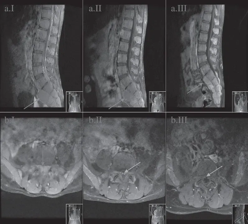
MRI of the lumbar spine in sagittal and axial views at the levels ofL5-S1. . A = sagittal view; B = axial view; I = initial presentation; II = day 14th after diagnosis; III = week 11th after diagnosis. The images show regression of the central spinal canal stenosis: from near-complete-obliteration on the initial image (B. II.1 red arrow) to the subsequent re-emergence of the spinal canal (B. II.2 and B. II.3 green arrow) with medical treatment. Also noted is the decrease in size of the bony masses and is the most prominent in the sacral region (II. A images blue arrow).
MRI of the lumbar spine in sagittal and axial views at the levels ofL5-S1. . A = sagittal view; B = axial view; I = initial presentation; II = day 14th after diagnosis; III = week…

The guided interpretation is available in the app

Epistemis explains what is seen in this image, why, and what it should not be confused with: guided findings, reasoned interpretation, and the clinical pearls of the case. Free; The image becomes available offline after downloading.

Download Epistemis

Epistemis is educational review material. It is not a medical device, does not diagnose or prescribe treatment, and does not replace formal medical training, current clinical guidelines, or professional clinical judgment.

Category
Thorax
Difficulty
advanced
Type
Radiological image
Mode
MRI

Origin: MultiCaRe · figure from open-access article PMC6116288 · License CC BY

Download Epistemis