
Epistemis

SPECT · Neuro / Head · open case 001

Radiology · Neuro / Head
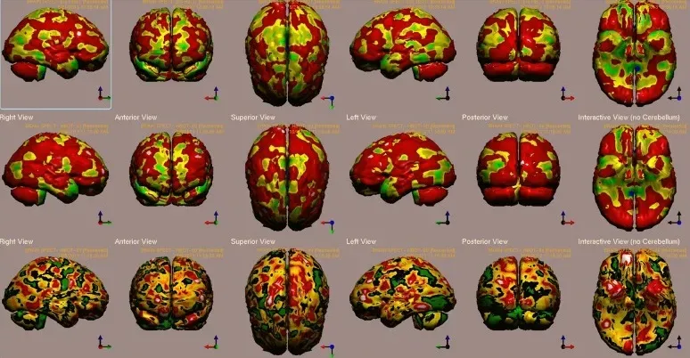
Case 1 single photon emission CT (SPECT) calculated change after hyperbaric oxygen therapy (HBOT) compared to baseline. In the Top two rows colors, represent maximal functional brain activity relative to the entire brain activity; in the Bottom row, colors represent regional changes in functional brain activity. White and red areas show the biggest relative changes in cerebral blood flow/perfusion. Significant changes can be seen at the right prefrontal and temporal areas (white). Abbreviations: Analysis of brain SPECT performed pre- and post HBOT showed the highest relative increase in activity (>15%) in the visual cortex (BA 19), prefrontal cortex (BA 9), orbito-frontal gyrus (BA 11), and paratemporal cortex (BA 37).
Case 1 single photon emission CT (SPECT) calculated change after hyperbaric oxygen therapy (HBOT) compared to baseline. In the Top two rows colors, represent maximal functional…

The guided interpretation is available in the app

Epistemis explains what is seen in this image, why, and what it should not be confused with: guided findings, reasoned interpretation, and the clinical pearls of the case. Free; The image becomes available offline after downloading.

Download Epistemis

Epistemis is educational review material. It is not a medical device, does not diagnose or prescribe treatment, and does not replace formal medical training, current clinical guidelines, or professional clinical judgment.

Category
Neuro / Head
Difficulty
advanced
Type
Radiological image
Mode
SPECT

Origin: MultiCaRe · figure from open-access article PMC5987035 · License CC BY

Download Epistemis